International Seminar-Workshop-Wellness through Ayurveda will be organized at Rishikesh on 3-4th October 2015..Please Register your self at http://ayushdarpan.org
Previous issues of AYUSH DARPAN in Hindi is now available online visit:http://ayushdarpan.org

मंगलवार, 25 जनवरी 2011

Hormonal disorders

Hormonal disorders are conditions that affect the endocrine system and the hormones they release. As such the effect of hormonal disorders can range throughout the whole body as different disorders can manifest in different areas. Hormone levels typically decrease with age and while some do not, hormone receptors become less active and cause certain changes to the body. However, simply replacing deficient hormones do not simply cure the problems caused and may even be harmful. Thus, treating hormonal disorders is not a simple task. Hormones are produced primarily in the endocrine glands but certain parts of the body such as the kidney or the placenta can also release hormones in conjunction with the endocrine system. Some glands in the body also produce not hormones but other substances such as the salivary gland and the sweat glands.

Typically hormone disorders involve either an overproduction by hyperactive glands or deficiency of a certain hormone by hypoactive glands. This may be due to a problem in the endocrine gland responsible for the production of the said hormone or the hypothalamus and the pituitary gland (sometimes referred to as the master gland) sending wrong signals to the body.

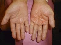
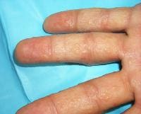
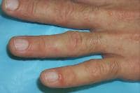
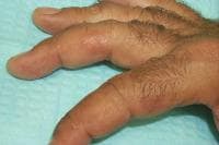
Impairment of normal bodily functions results from hormonal disorder. For example, those suffering from diabetes suffer from a low production or poor reception of insulin, a hormone responsible for metabolizing sugars. Thus they experience elevated blood sugar levels. Overproduction of the growth hormone from the pituitary glands causes a massive and sometimes uncontrollable growth spurt, causing individuals to be abnormally tall. This condition is known as gigantism. Thyroid disorders can produce a variety of different conditions from metabolic problems to blood pressure issues. Some hormones such as prolactin can even stimulate the growth of tumors, such as breast cancer. Thus depending on the hormonal disorder, the manifested symptoms may vary greatly. Some may not even manifest symptoms immediately until the problem is severe. A blood test may help the doctor check hormone levels in the blood, or the doctor may ask you to perform certain actions or take certain food or medicine to stimulate hormone production and observe its effects. Examples of this treatment include consumption of sugary foods to induce insulin production, or asking the patient to fast.

There are many different causes of hormonal disorders. Some may be genetically linked, as some may seem to run within the family, such as diabetes. Others may involve nutritional factors. Goiter, the condition of enlargement of the thyroid gland, is related to a lack of iodine in the diet, while diabetes, a lack of the hormone insulin or the body’s poor reception to it, can be aggravated by a sugar and carbohydrate-rich diet. Age also plays a factor in the development of hormonal disorders, especially in the production of sex hormones which reduces in both sexes as they age. This is the reason why women experience menopause. Injury to the hormone producing organs can also cause inhibition or overstimulation of hormone production.

Sometimes autoimmune disorders can affect the hormone production by attacking hormone production sites in the body, disrupting the normal processes of the organ. Tumors can also grow on hormone producing organs and cause an overproduction of the hormone or stifle hormone producing tissue in their growth, causing a decrease in hormone production. Other factors that may affect hormone production include stress, infection and changes in your blood chemistry and electrolyte balance.

Problems in the pituitary gland in particular can cause a wide range of other hormonal disorders, since it controls most of the other glands in the body such as thyroid-stimulating hormone deficiency.

Recent studies also show that certain chemicals described as “endocrine disruptors” play a part in causing hormonal disorders. These endocrine disruptors may mimic the effects or slightly mimic the effects of naturally-occurring hormones, causing an overstimulation of processes. Some may bind to the hormone receptors located in different parts of the body. When the body normally produces hormones, the receptors fail to pick up the signal and thus the body cannot respond properly. Others may disrupt the production of the hormones themselves, or the action of the receptors. These endocrine disruptors may find its way in daily household objects, cosmetics and even our food. Some evidence has shown that these endocrine disruptors may also change the expression of DNA and their effects may be passed on to the next generation.

Hormone replacement therapy is a popular treatment to many types of hormonal disorders. Hormonal supplements may come in oral or intravenous form. However, not all hormonal disorders may be treated with hormone replacement therapy. An example is in women who experience menopause, where hormone replacement therapy may increase risks in certain cancers and cardiovascular diseases. Others such depend on maintenance drugs that mimic the hormone’s action or increase the body’s reception to the hormone. Supplements may also help restore normal hormone development as some may be caused by deficiencies in certain vitamins or minerals. In extreme cases of hormone over production, surgical means may be used to treat the over production. Corrective surgery to the involved hormone producing sites may be used to stem the production of hormones, such as in the pituitary gland in cases of gigantism. Certain drugs can also block the production of overproduced hormones or bind to their receptors to reduce their effect.

If hormonal disorders prevent proper balance of body chemistry or the metabolizing of certain nutrients, then an adjustment in diet may be helpful in lessening the pressure on the body. This is particularly useful in diabetes and thyroid conditions. Exercise may also help release some hormones as well as help facilitate metabolic processes. Proper treatment of individual disorders should be facilitated by the doctor based on the patient’s condition, medical history, and other personal details.

Sarcoidosis


Sarcoidosis is a disease that features a specific type of inflammation of various tissues of the body. Sarcoidosis can appear in almost any body organ, but it starts most often in the lungs or lymph nodes. As sarcoidosis progresses, microscopiclumps of a specific form of inflammation, called granulomas, appear in the affected tissues. In the majority of cases, these granulomas clear up, either with or without treatment. In the few cases in which the granulomas do not heal and disappear, the tissues involved tend to remain inflamed and become scarred (fibrotic). In addition to the lungs and lymph nodes, the organs more likely than others to be affected by sarcoidosis are the liver, skin, heart, nervous system, and kidneys, in that order of frequency.
Sarcoidosis is sometimes named according to the organ involved.
  • When sarcoidosis affects the lungs, it can be referred to as lung sarcoidosis or pulmonary sarcoidosis.
  • When sarcoidosis affects the liver, it can be referred to as hepatic sarcoidosis.
  • When sarcoidosis affects the skin, it can be referred to as skin sarcoidosis or sarcoid dermatitis.
  • When sarcoidosis affects the heart, it can be referred to as heart sarcoidosis or cardiac sarcoidosis.
  • When sarcoidosis affects the nervous system including the brain, it can be referred to as neurological sarcoidosis or neurosarcoidosis.
  • When sarcoidosis affects the kidneys, it can be referred to as kidney sarcoidosis or renal sarcoidosis.

Sarcoidosis Causes

The cause of sarcoidosis is unknown. Sarcoidosis is currently thought to be associated with an abnormal immune response. It is not known whether the triggerthat initiates the immune disturbance is a foreign substance, chemical, drug, virus, or some other substance. Sarcoidosis is not acancer. It is not contagious, and friends and family will not catch it from an affected individual. Although it can occur in families, there is no evidence that sarcoidosis is passed from parents to children.

Sarcoidosis Symptoms

Sarcoidosis can appear suddenly and disappear. Alternatively, it can develop gradually and go on to produce symptoms that come and go, sometimes for a lifetime.
The symptoms of sarcoidosis depend on what areas of the body are affected.
  • Shortness of breath (dyspnea) and a coughthat won't go away can be among the first symptoms of sarcoidosis.
  • But sarcoidosis can also show up suddenly with the appearance of skin rashes.

    • Tender, raised, red bumps (called subcutaneous sarcoidosis or erythema nodosum) on the shins of the legs, or less frequently on the arms, are common and can cause leg or arm pain.
    • A rash on the surface of the skin (cutaneous sarcoidosis or sarcoid dermatitis) of the face occurs frequently.
  • Inflammation of the eyes can also occur.
More generalized symptoms of sarcoidosis include:
  • weight loss,
  • fatigue,
  • night sweats,
  • fever, or
  • just an overall feeling of ill health.
It is important to note that sarcoidosis is usually not crippling. It often goes away by itself, often healing in 24 to 36 months. Even when sarcoidosis lasts longer, most patients can go about their lives as usual

When to Seek Medical Care

Anyone with the symptoms of shortness of breath and persistent cough should have an evaluation by a health care professional. Furthermore, those with a persistent rash, weight loss, fatigue, night sweats, and/or fever should have a medical examination. Moreover, patients with a known diagnosis of sarcoidosis should have medical follow-up.

Exams and Tests

The preliminary diagnosis of sarcoidosis is based on the patient's medical history, routine tests, a physical examination, and a chest X-ray. The doctor confirms the diagnosis of sarcoidosis by eliminating other diseases with similar features.
These include such granulomatous diseases:
  • as berylliosis (a disease resulting from exposure to beryllium metal),
  • tuberculosis, farmer's lung disease (hypersensitivity pneumonitis),
  • fungal infections,
  • rheumatoid arthritis,
  • rheumatic fever, and
  • cancer of the lymph nodes (lymphoma).
No single test can be relied on for a correct diagnosis of sarcoidosis. X-rays and blood tests are usually the first procedures the doctor will order. Pulmonary function tests often provide clues to diagnosis. Other tests may also be used, some more often than others.
The biopsy of a tissue sample of an involved organ is the ultimate test to confirm the diagnosis. Many of the tests that the doctor uses to help diagnose sarcoidosis can also help the doctor follow the progress of the disease, and determine whether the sarcoidosis is getting better or worse. The following are commonly used tests in the evaluation of a patient with sarcoidosis.
Chest X-ray
The chest X-ray is often helpful to give the doctor a picture of the lungs, heart, as well as the surrounding tissues containing lymph nodes (where infection-fighting white blood cells form) and give the first indication of sarcoidosis. For example, a swelling of the lymph glands between the lungs can show up on an X-ray. An X-ray can also show which areas of the lung are affected.
Pulmonary function tests
By performing a variety of tests called pulmonary function tests (PFTs), the doctor can find out how well the lungs are doing their job of expanding and exchangingoxygen and carbon dioxide with the blood. The lungs of patients with sarcoidosis cannot handle these tasks as well as they should; this is because granulomas and fibrosis of lung tissue decrease lung capacity and disturb the normal flow of gases between the lungs and the blood. One PFT procedure calls for the patient to breathe into a machine called a spirometer. It is a mechanical device that records changes in the lung size as air is inhaled and exhaled, as well as the time it takes the patient to do this.
Blood tests
Blood analyses can evaluate the number and types of blood cells in the body and how well the cells are functioning. They can also measure the levels of various blood proteins known to be involved in immunological activities, and they can show increases in serum calcium levels and abnormal liver function that often accompany sarcoidosis.
Blood tests can measure a blood substance called angiotensin converting enzyme (ACE). Because the cells that make up granulomas secrete large amounts of ACE, these enzyme levels are often high in patients with sarcoidosis. Blood ACE levels, however, are not always elevated in people with sarcoidosis, and increased ACE levels can also occur in other illnesses.
Bronchoalveolar lavage
This test uses an instrument called a bronchoscope - a long, narrow tube with a light at the end - to wash out, or lavage, cells and other materials from inside the lungs. This wash fluid is then examined for the amount of various cells and other substances that reflect inflammation and immune activity in the lungs. A high number of white blood cells in this fluid usually indicates an inflammation in the lungs.
Biopsy
Microscopic examination of specimens of lung tissue obtained with a bronchoscope, or of specimens from other tissues, can tell a doctor where granulomas have formed in the body and can provide the ultimate diagnosis.
Gallium scanning
In this procedure, the doctor injects the radioactive chemical element gallium-67 into the patient's vein. The gallium collects at places in the body affected by sarcoidosis and other inflammatory conditions. Two days after the injection, the body is scanned for radioactivity. Increases in gallium uptake at any site in the body indicate that inflammatory activity has developed at the site and give an idea of which tissue, and how much tissue, has been affected. However, since any type of inflammation causes gallium uptake, a positive gallium scan does not necessarily mean that the patient has sarcoidosis.
Slit-lamp examination
An instrument called a slit lamp, which permits examination of the inside of the eye, can be used to detect silent eye damage from sarcoidosis.


शनिवार, 22 जनवरी 2011

Medicinal values of Betel Leaf


Betel-LeafThe Betel (Piper betle) is the leaf of a vine belonging to the Piperaceae family. The plant, by itself, is said to have originated in India. Betel Leaf is cultivated in majority of South and Southeast Asia, and is a much esteemed leaf across the dozen nations. In India, the leaf is considered auspicious and is used as an auspicious exchange material not only during ceremonies, but also when fixing deals, business transactions and even marriage alliances.
Although the betel leaf earned a bad reputation during mid-1980s when early Western literature suggested that the betel liquid causes oral cancer, Dr. S. V. Bhide and others at the Cancer Institute in India, showed that it is not the leaf, but some contents of the areca nut (areca nut, lime and tobacco are chewed with betel leaf) and the tobacco that are the culprits.
The betel leaf when taken alone (without the said accompaniments), has several medicinal benefits, which very few of are aware of.
Discussed here are the less-known medicinal properties of betel leaf. Apart from being a mild stimulant, betel leaf is used for various medicinal purposes.
To begin with, when the leaf is chewed, the mild anti-infective content in the leaf, freshen breath, and cleanses the mouth. Its constituents enter the blood directly via the buccal mucosa. As this is a direct way of entry into blood stream, it is the best way to deliver drugs into the blood stream during sickness.
On chewing, the betel leaf induces salivation. Saliva fights bacteria in the mouth, and helps reduce plaque formation on teeth. Further, the Betel leaf contains compounds that act as heart beat regulators, relaxing the blood vessels.
The leaf contains certain poly-phenols that not only fight microbes, but also act as pain relievers and anti-inflammatory agents. Recent studies have shown that the leaf contains tannins, sugar, diastases and an essential oil. A particular phenol called ‘chavicol’, present in it has powerful antiseptic properties.
The leaf has analgesic and cooling properties, and can be applied on the painful area for temporary relief.
Betel leaves possess good diuretic properties. Therefore, it can be mixed with dilute milk and consumed by sweetening, as it helps ease urination.
The betel leaves are also beneficial in treating nervous pains and debility. The juice of the leaves, when consumed by adding a teaspoon of honey, twice a day, acts a good tonic.
Betel leaf has been in use since ancient times for healing wounds. Ayurveda has strongly believed in this property of the leaf. The juices of the leaves are applied locally on the wound, after which, the betel leaf is wrapped around and bandaged. The wound is sure to heal within a day or two.
Also, recent studies have shown that the leaf contains components that have chemo-preventive and anti-cancer properties.
In short, Betel leaf is a storehouse of several chemicals that are of much pharmaceutical value. Studies are on, to isolate and identify each molecule and study their effects. It is hoped that several more medicinal properties of this wonderful leaf are yet to be revealed. However, the one precaution to be taken is to avoid chewing the leaf with areca nut and tobacco, instead, try some variety spices including cloves, cardamom, cinnamon and the likes to enjoy its benefits.
Related posts:
  1. Betel Leaf (Piper Betel) promotes good health!!
  2. Medicinal and nutritive values of the ‘Bael’
  3. Olive leaf extract can help tackle obesity
  4. Medicinal values of herbs and spices
  5. Caraway has medicinal values too!!

Dyshidrotic Eczema


Background

Dyshidrotic eczema is a recurrent or chronic relapsing form of vesicular palmoplantar dermatitis of unknown etiology. Dyshidrotic eczema also is termed pompholyx, which derives fromcheiropompholyx, which means "hand and bubble" in Greek.

The etiology of dyshidrotic eczema is unresolved and is believed to be multifactorial. Dyshidrotic eczema is considered a reaction pattern caused by various endogenous conditions and exogenous factors.

Pathophysiology

The hypothesis of sweat gland dysfunction has been disputed because vesicular lesions have not been shown to be associated with sweat ducts. A 2009 case report provided clear histopathologic evidence that sweat glands do not play a role in dyshidrosis.1 However, hyperhidrosis is an aggravating factor in 40% of patients with dyshidrotic eczema. Improvement in pruritus, erythema, vesicles, and hand dermatitis with fewer or no signs of relapse has been obtained after botulinum toxin A injection.2 

Dyshidrotic eczema may be associated with atopy and familial atopy. Of patients with dyshidrosis, 50% have atopic dermatitis.

Exogenous factors (eg, contact dermatitis to nickel, balsam, cobalt; sensitivity to ingested metals; dermatophyte infection; bacterial infection) may trigger episodes. These antigens may act as haptens with a specific affinity for palmoplantar proteins of the stratum lucidum of the epidermis. The binding of these haptens to tissue receptor sites may initiate pompholyx.

Evidence shows that the ingestion of metal ions such as cobalt can induce both type I and type IV hypersensitivity reactions, and, in addition, they can also act as atypical haptens activating T lymphocytes through human leucocyte antigen–independent pathways, causing systemic allergic dermatitis in the form of dyshidrotic eczema.3,4 

Emotional stress5 and environmental factors (eg, seasonal changes, hot or cold temperatures, humidity) reportedly exacerbate dyshidrosis.

Dyshidrosis-like eczematous eruptions with the use of intravenous immunoglobulin infusions have been reported.

In some patients, a distant fungal infection can cause palmar pompholyx as an id reaction. In one study, one third of pompholyx occurrences on the palms resolved after treatment for tinea pedis.

Frequency

United States

Dyshidrotic eczema occurs in 5-20% of patients with hand eczema and more commonly occurs in warmer climates and during spring and summer months (seasonal or summer pompholyx).

International

Dyshidrotic eczema comprised 1% of initial consultations in a 1-year Swedish study. In a study of 107,206 Swedish individuals, 51 (0.05%) were diagnosed with dyshidrosis. Of all hand dermatitis cases in that population, 3% had dyshidrosis.6 

In a retrospective study reviewing records of 714 Portuguese patients during a 6-year period, Magina et al found dyshidrotic eczema to be the third most common type of hand dermatitis (20.3%).7

Mortality/Morbidity

Dyshidrotic eczema can be severe, resulting in occupational disability and time away from work; however, disability compensation usually is not provided for this condition.

Sex

The male-to-female ratio for dyshidrotic eczema is 1:1.

Age

Dyshidrotic eczema affects individuals aged 4-76 years; the mean age is 38 years. After middle age, the frequency of dyshidrotic eczema episodes tends to decrease.

Clinical

History

Patients report pruritus of hands and feet with a sudden onset of vesicles. Burning pain or pruritus occasionally may be experienced before vesicles appear. Dyshidrotic eczema episodes vary in frequency from once per month to once per year. Patients with dyshidrotic eczema may report a variety of factors that possibly are related to eruptions, as follows:
  • Emotional stress
  • Personal or familial atopic diathesis (eg, asthma, hay fever, sinusitis)
  • Certain work exposures (eg, cobalt) and/or recreational exposures
  • Recent exposure to contact allergens (eg, nickel, balsams, paraphenylenediamine, chromate, sesquiterpene lactones) before condition flares
  • Exposure to contact irritants before condition flares
  • Recent exposure to costume jewelry (patients with palmar pompholyx and allergic to nickel)
  • Recent treatment with intravenous immunoglobulin therapy8,9
  • HIV related: Two cases were reported of HIV-positive patients who developed dyshidrotic eczema as an immune reconstitution inflammatory syndrome shortly after highly active antiretroviral therapy.10 Pompholyx has also been described as a manifestation of symptomatic HIV infection, including individuals who do not respond to topical and systemic therapies and whose condition resolves only after initiation of combination antiretroviral therapy.11

Physical

Symmetric crops of clear vesicles and/or bullae on the palms and lateral aspects of fingers characterize dyshidrotic eczema (see the images below). Feet, soles, and the lateral aspects of toes also may be affected.
Tense vesicles and bullae on the palm. Courtesy o...

Tense vesicles and bullae on the palm. Courtesy of Norman Minars, MD, University of Miami, Department of Dermatology & Cutaneous Surgery.


Small tense vesicles on the fingers.

Small tense vesicles on the fingers.


Small, discrete, coalesced vesicles on the dorsal...

Small, discrete, coalesced vesicles on the dorsal hand.


Small, discrete, coalesced vesicles on the finger...

Small, discrete, coalesced vesicles on the fingers.


In mildly affected patients, vesicles are present only on the lateral aspects of the fingers and, occasionally, involve feet and toes (see image below).

Small discrete vesicles of the lateral fingers.

Small discrete vesicles of the lateral fingers.


Vesicles are deep seated with a tapiocalike appearance, without surrounding erythema. They may become large, form bullae, and become confluent. Vesicles typically resolve without rupturing, followed by desquamation. See image below.
Close-up view of tense vesicles and bullae of the...

Close-up view of tense vesicles and bullae of the palm. Courtesy of Norman Minars, MD, University of Miami, Department of Dermatology & Cutaneous Surgery.


Hands are involved solely in 80% of patients, feet solely in 10%), and both hands and feet are involved in 10% of patients. See the images below.
Discrete yellow pustules on the sole of the foot....

Discrete yellow pustules on the sole of the foot. Courtesy of Norman Minars, MD, University of Miami, Department of Dermatology & Cutaneous Surgery.


Palms and soles of a patient with a dyshidrosis f...

Palms and soles of a patient with a dyshidrosis flare. The patient unroofed a large bulla on the right sole.


With long-standing disease, patients' fingernails may reveal dystrophic changes (eg, irregular transverse ridging, pitting, thickening, discoloration). Interdigital maceration and desquamation of the interdigital spaces often are present, despite the possible absence of a dermatophyte infection. Vesicles and/or bullae may become infected secondarily, and pustular lesions may be present. Cellulitis and lymphangitis may develop.
The Dyshidrotic Eczema Area and Severity Index was developed based on severity grades for the number of vesicles per square centimeter, erythema, desquamation, itch, and the extent of affected areas.12 The index was found to be a simple standardized method for assessing the condition and was used to assess disease severity and treatment effectiveness in 2 clinical studies. Further evaluation with larger patient groups is needed.

Causes

The cause of dyshidrotic eczema is unknown. The condition often appears related to other skin diseases (eg, atopic dermatitis, contact dermatitis, allergy to ingested metals, dermatophyte infection, bacterial infection, environmental or emotional stress). Several factors may participate in causing dyshidrotic eczema, as follows:
  • Genetic factors: Monozygotic twins have been affected simultaneously by dyshidrotic eczema. The pompholyx gene has been mapped to band 18q22.1-18q22.3, in the autosomal dominant form of familial pompholyx.13
  • Atopy: As many as 50% of patients with dyshidrotic eczema have reportedly had personal or familial atopic diathesis (eczema, asthma, hayfever, allergic sinusitis). The serum immunoglobulin E (IgE) level frequently is increased, even in patients who do not report a personal or familial history of atopy. Occasionally, dyshidrotic eczema is the first manifestation of an atopic diathesis.
  • Nickel sensitivity: This may be a significant factor in dyshidrotic eczema. Nickel sensitivity was reportedly low in some studies of dyshidrosis patients but significantly elevated in other studies. Increased nickel excretion in the urine has been reported during exacerbations of pompholyx. Ingested metals have been found to provoke exacerbations of pompholyx in some patients.
  • Low-nickel diets: These have reportedly decreased the frequency and severity of pompholyx flares. A high palmoplantar perspiration rate has been suggested to result in a local concentration of metal salts that may provoke the vesicular reaction. Contact allergy has been documented in 30% of patients with dyshidrotic eczema.
  • Cobalt sensitivity: The oral ingestion of cobalt manifests systemic allergic dermatitis as dyshidrotic eczema less frequently than the oral ingestion of nickel. Much more common is the simultaneous occurrence nickel and cobalt allergy seen in 25% of nickel-sensitive patients developing pompholyx. In these cases, the eczema is usually more severe. When suspected as the cause of the dyshidrotic eczema, high oral ingestion of cobalt should be taken in consideration, regardless of the patch test results.3
  • Low-cobalt diet: A point-based diet has been proposed to help patients limit cobalt ingestion and to keep the serum level below the threshold for developing flares, which is approximately less than 12 mcg/d. This diet has demonstrated higher compliance than an avoidance diet list. In addition, this diet also reduces the amount of nickel consumed.3
  • Exposure to sensitizing chemicals or metals: Dyshidrotic eczema outbreaks are sometimes associated with exposure to sensitizing chemicals or metals (eg, chromium, cobalt, carba mix, fragrance mix, diaminodiphenylmethane, dichromates, benzoisothiazolones, paraphenylenediamine, perfumes, fragrances, balsam of Peru, Primula plant).
  • Id reaction: Controversy surrounds the possible existence of an id reaction, which is considered to be a distant dermatophyte infection (tinea pedis, kerion of scalp) triggering a palmar pompholyx reaction (also termed pompholyx dermatophytid).
  • Fungal infection: Pompholyx occasionally resolves when a tinea pedis infection is treated, then relapses when the fungal infection recurs, supporting the existence of this reaction pattern. Of patients who have a vesicular reaction to intradermal trichophytin testing, less than one third have experienced a resolution of pompholyx after treatment with antifungal agents.
  • Emotional stress: This is a possible factor in dyshidrotic eczema. Many patients report recurrences of pompholyx during stressful periods. Improvement of dyshidrotic eczema using biofeedback techniques for stress reduction supports this hypothesis.
  • UV-A light: A group of 5 patients with pompholyx developed typical lesions morphologically and histologically consistent with a vesicular dermatitis after a provocation with long-wavelength UV-A light. Of interest, UV-B phototherapy and photochemotherapy are well-known efficient therapies for pompholyx. Further studies ruled out contact dermatitis, polymorphic light eruption, and heat as the culprit of the lesions in these patients, confirming that the reaction was a true photosensitivity rather than a photoaggravation.14 Pompholyx caused by exposure to UV-A may be considered a variation of seasonal (summer) pompholyx.
  • Other factors: Isolated reports describe other possible causative factors, such as aspirin ingestion, oral contraceptives, cigarette smoking, and implanted metals, among others. A 3-year prospective study of the causes of dyshidrotic eczema (pompholyx) in 120 patients found causes of pompholyx related to contact exposure (67.5%), including cosmetic products (31.7%) and metals (16.7%); interdigital-plantar intertrigo (10%); and internal causes (6.7%), with an additional 15% with undiagnosed (idiopathic) causes, probably related to atopic factors.15 Specifically, note the following:
    • Contact allergy was found in 89 (74.2%) of 120 of the patients. The most frequent allergens were nickel, shower gel, chromium, fragrance, shampoo, and balsam of Peru. Less frequent allergens were lanolin, cobalt, thiuram, lauryl sulfate, fresh tobacco, p -phenylenediamine (PPD), formaldehyde, parabens, and octyl gallate. In 97 of 193 positive patch test results, correlation existed between the application of the agent and pompholyx recurrence. The relevance of the analysis was confirmed in 81 (67.5%) of 120 patients. In summary, the most frequent causes of pompholyx related to contact with substances were hygiene product intolerance (46.7%), metal allergy (25%), and others (28.3%).
    • Intertrigo occurred in 19 (15.8%) of 120 patients. Of those, 80% presented with dermatophytosis and 20% presented with candidiasis. After 3 weeks of antifungal therapy, 6 of 19 patients remained symptomatic for pompholyx.
    • With regard to internal causes, 30 patients presented with a positive patch test result for metals, but only 2 presented with exacerbations of the lesions after a challenge test.
    • Of 58 patients with a history of smoking tobacco, 5 presented with a positive reaction, and 2 of those reactions were considered relevant.
    • Drug allergy was determined to be the causative agent in 3 patients (amoxicillin in 2 and intravenous immunoglobulin in 1).
    • Food-related pompholyx was detected in 4 patients, and after a challenge test, reactivation occurred in 3 patients (2 for paprika and 1 for orange juice).

शुक्रवार, 21 जनवरी 2011

Erection Problem


Sankalpapravano nityam priyaam vashyaamapi sthreeyam I
na yaathi lingashaithilyaath kadaachidyaathi vaa yadi II
Shwaasaarthaha swinnagaatrshcha moghasankalpacheshtitaha I
mlaanashishnashcha nirbeejaha syodetat klaibyalaxanam II
Even though a man has a strong desire to perform sexual act with a cooperative partner, he cannot perform sexual act because of looseness or absence of erection of his penis. Even if he performs sexual act with his determined efforts he does not get erection and gets afflicted with tiredness, perspiration and frustration to perform sex.
Erectile dysfunction (ED) thus is the inability of a male to achieve or maintain an erection sufficient for intercourse.
Erectile dysfunction may sometimes be even termed as “impotence”. The term "erectile dysfunction" can mean the inability to achieve erection, an inconsistent ability to do so, or the ability to achieve only brief erections.
As a general estimate 2-4 out of 10 males suffer from this problem chronically and there are many males who suffer from this problem time to time! Until recently, doctors thought impotence was mainly due to psychological causes. But in the recent year doctors have started believing more than half of all the cases are due to by physical problems.
Causes of ED
1. According to Ayurveda the diseases which cause erectile dysfunction are –
  • · Heart diseases
  • · Anemia
  • · Asthma
  • · Liver disorders
  • · Tiredness
2. Stress, Depression, Arteriosclerosis (hardening of the arteries) causes erection problems.
3. So many other Physiological or even psychological or some addiction like smoking and alcoholism related factor are responsible for Erection Problem.
4. Diabetes is one of the main cause amount 25 % of the patients suffering from ED as diabetes may cause nerve deterioration.
5. Dry food, drinks and medicines" cause impotence or erectile dysfunction.
6. Apart from these the imbalance in tridoshas also causes impotence or erectile dysfunction.
How to check erection problem?
Almost all men fail to get an erection some time or the other. This is a normal. However, if the erectile dysfunction seems to bother 25% of the time then one should take medical help especially ayurvedic consultation as it might be a case of male impotence. Men who face Erection Problem must know that they should not neglect the persisting problem and must consult a doctor as soon as possible rather than wait passively.
Remedies for Erectile Dysfunction:
Erectile dysfunction is treatable at any age. In Ayurveda; total treatment in for impotence is called as “Vaajikarana therapy”. This therapy increases the strength of a man to perform sexual act, like a horse.
Vaajikarana therapy leads to-
  • Happiness in life.
  • Good strength in male sexual organ i.e.Penis.
  • Potency to produce child.
  • Increased span/stamina of erection. 
Who is Eligible for Vaajikarana Therapy ?
The vajikarana therapy should be administered to persons who are between 18 to 70 years of age.
Which Ayurvedic Treatment may help you? There are so many treatments and products available in Ayurveda to treat Impotence (ED) of Men. With our Ayurveda formulations you do not have to live with this problem. In most cases, impotence can be successfully treated by our Ayurveda Treatments and patients have been leading their normal happy sexual life.


Vd.Sushant S Patil
Vidyanagar, bhusawal road,
Faizpur425503
Tal-yawal dist-jalgaon
  9860431004

Kumkumadi Lepam- An ayurveda beauty preparation


Kumkumadi lepam is an age old ayurveda remedy for acne and pimples ( known as youvana pitikas in ayurveda), dark circles,pigments, scars and stretch marks. Kumkumadi lepam contains skin friendly herbs like Saffron, sandal wood and Turmeric . 


Action of Ingredients:

Saffron: Saffron helps to reduce dark pigments, dark circles under eyes, acne and pimple. These magic red stigmas increase fairnessand glow of skin when applied as face pack.

Sandal wood: sandalwood has many antimicrobial properties. Ayurveda acharyas recommend use of sandal wood as a part of natural skin care regimes to heal and soothe many skin ailments and conditions. One of the primary uses of this wonder herb is that it can kill germs, soothe inflamed skin, and clear blemishes left even by stubborn acne. Regular use of sandalwood helps to get clean and clearskin.

Turmeric: Turmeric nourishes skin and gives it a healthy natural glow and radiance. It reduces inflammation of skin, soothens it and prevents many skin ailments. The golden yellow coloured turmeric powder acts as natural antibiotic and helps to fight acne and pimple. Texts of ayurveda praise the scar and blemish reducing properties of turmeric. Continuous use of turmeric reduces stretch marks and gives the natural tone for skin.

Kumkumadi lepam is natural cream which has multiple uses. It contains all the above herbs as active ingredients. Kumkumadi lepam is only for external use.

Acne and pimples: Kumkumadi lepam helps to reduce acne andpimples which erupt due to dry skin. Kumkumadi lepam has to be applied on affected area after cleansing the skin .Neem tulsi soapcleanses the skin perfectly by removing dirt and acts as natural antibiotic too. Saffron in kumkumadi lepam is known for its healing properties and also it imparts glow to skin. Thus saffron heals acne, while turmeric fights with germs and sandal wood soothes the skin which is irritated due to microbial attack.

Scars and Blemish: Kumkumadi lepam is known to reduce scars and blemish from ages. Ayurveda acharyas eulogize the scar and blemish removing properties of kumkumadi lepam. Little of this cream has to be rubbed on affected area at night and washed off next day morning with warm water and mild soap like Neem tulsi soap.

Stretch Marks: Stretch marks spoil the beauty of skin. Kumkumadi lepam helps to reduce stretch marks and gives an even tone to skin. Regular use of kumkumadi lepam is useful in stretch marks due to pregnancy, obesity, muscle building etc.

Dark circles and pigments: Kumkumadi lepam is also advised by ayurveda practitioners as an effective remedy in dark circles andpigments. Regular use of this preparation help to enhance beauty of skin by reducing dark circles and pigments.

This article is copy righted. The author Dr.SavithaSuri is an Ayurvedic Physician and web master of Ayurveda help through ayurveda remedies .Read more about kukumadi lepam atkumkumadi lepam
http://ayur-help.blogspot.com/search/label/ayurveda

Psoriasis


Psoriasis

Psoriasis is a chronic skin disease, in which the skin surface covers with dark red scaly patches. People between the age group 14 and 32 years are more prone to this disease. Psoriasis generally attacks on genital and surrounded area, underarms, scalp, elbows, and ear. Psoriasis can be caused due to many reasons. It can be due to infection or may be due to stress. The extent of psoriasis varies from person to person. Some patients have psoriasis in selective parts of their body, while some have their entire body affected due to this. Psoriasis attacks specially those who consume lots of junk food, has w eak metabolic activity, and suffer from bacterial infections. It also attacks those who live a stressful life or suffer from high blood pressure. In ayurveda psoriasis is known as kitabh, a disease that is included in kushth roga. This article will explain different aspects of psoriasis, its cause, symptoms and ayurvedictreatments/home remedies.

Psoriasis Causes

The major psoriasis causes are skin infection, stressful lifestyle, over emotional nature. Various researches also proved that there are certain blood cells in our body which when become impulsive and hyperactive cause psoriasis. These blood cells cause allergy to the skin and also cause other skin-related diseases. There are people who cannot adjust in certain temperature and climate, and suffer from skin disease like psoriasis. Psoriasis can also be a side effect or reaction of the consumption of strong drugs and medicine. People suffering from asthma, poor digestive system, weak immune system, also suffer from psoriasis.

Psoriasis Diet

Diet rich in green vegetables like cucumber, carrot, etc., fruits, are good for psoriasis patient. It is also found that patient who lives on fruit-based diets for a month experiences much better result than any other medicine. Common fruits used in the diet are beets, grapes, cucumber, etc. Once the fruit-based diet is over, go for balanced diet containing cereals, grams, green leafy vegetables, juicy fruits, etc. Also include cheese, curd, etc. Junk food, oily and spicy foods should be strictly avoided. Milk products, egg, caffeine, chocolates, sugar, etc., are a big no for psoriasis patient.
Taking bath in sea water is very good for the psoriasis treatment. Similarly, taking bath in Epsom salt or oatmeal containing water is also good. Remember do not use chemical-based soaps as it will make the problem severe and painful. Another thing is do not bath too much, bathing 3-4 times a day. Always dry your body with soft, clean towel after bath.
Similarly, gentle massage over the infected area using avocado or cashewnut oil is very good for the treatment of psoriasis.
Band gobhi (Cabbage)
Green leafy cabbage is one of the best home remedy for psoriasis. This is used as a covering for the infected area. Take the outermost layer of a cabbage, clean it using warm water. Rinse all the extra water by pressing between a towel. Once the leaf is dried, spread it on the slab, and flatten it by pressing with hand or any other thing available at home. Warm the leaf and gently cover it over the infected area. Cover it with a clean cloth. This process should be repeated for 3-4 times a day for the period when the infected area is not fully recovered.
Apart from the above mentioned home remedies, given below are some of the other important things that can be done to get relief from psoriasis. Give your body enough sunlight, i.e., Vitamin D, however, not excess. Always use clean soft towel to dry your body completely after bath, use good quality moisturizer, there are many natural herb based moisturizer available in the market. However, you may also prepare some moisturizer at your home.

Psoriasis Symptoms

Patient suffering from psoriasis experiences severe itching in the skin, dark red patches over and around the infected skin area. Patient may feel stressful, irritated, and helpless. Psoriasis patient also experiences headache, weak memory, bad body smell, etc.

Home Remedies for Psoriasis - Psoriasis Treatment

Given below are some of the effective and useful home remedies for the psoriasis treatment. However, it is not necessary that all these below mentioned remedies will be effective for every psoriasis patient.

Dahi (Curd)

Make fresh glass of buttermilk from curd and drink 2-3 times a day. Buttermilk is a healthy drink for psoriasis patient. It also cleans the toxic substance within the stomach and helps in digestion. This is one of the useful home remedies for psoriasis.

Vitamin E-rich food

It is also found that Vitamin E-rich food is good for patients suffering from psoriasis. Some of the Vitamin E-rich food is spinach, almond oil, sweet potato, sunflower seeds, and wheat germs.

Karela (Bitter gourd) Juice

Juice extracted from bitter gourd is an effective home remedy for the psoriasis treatment. Patient suffering from psoriasis should take 1-2 cup of bitter gourd juice in empty stomach in the morning. Continue this for 5-6 months. Patient may add a tablespoon of lime juice, if found difficult to digest the bitter taste. This is one of the best home remedies for psoriasis.

Lecithin seeds

Lecithin seeds are another effective home remedy for the treatment of psoriasis. Take 3-4 tablespoon of lecithin seeds every day for 2-3 month. This has a magical effect and shows great result within 2-3 months.

(Til) Sesame seeds

Sesame seeds are good for the treatment of various skin-related diseases. Take 15-20 sesame seeds and soak in a glass of water. Keep it overnight and drink in empty stomach early in the morning. This is one of the important home remedies for psoriasis.
Other vegetable seeds important for the psoriasis treatment are sunflower and pumpkin seeds.