Background
Dyshidrotic eczema is a recurrent or chronic relapsing form of vesicular palmoplantar dermatitis of unknown etiology. Dyshidrotic eczema also is termed pompholyx, which derives fromcheiropompholyx, which means "hand and bubble" in Greek.
The etiology of dyshidrotic eczema is unresolved and is believed to be multifactorial. Dyshidrotic eczema is considered a reaction pattern caused by various endogenous conditions and exogenous factors.
The etiology of dyshidrotic eczema is unresolved and is believed to be multifactorial. Dyshidrotic eczema is considered a reaction pattern caused by various endogenous conditions and exogenous factors.
Pathophysiology
The hypothesis of sweat gland dysfunction has been disputed because vesicular lesions have not been shown to be associated with sweat ducts. A 2009 case report provided clear histopathologic evidence that sweat glands do not play a role in dyshidrosis.1 However, hyperhidrosis is an aggravating factor in 40% of patients with dyshidrotic eczema. Improvement in pruritus, erythema, vesicles, and hand dermatitis with fewer or no signs of relapse has been obtained after botulinum toxin A injection.2
Dyshidrotic eczema may be associated with atopy and familial atopy. Of patients with dyshidrosis, 50% have atopic dermatitis.
Exogenous factors (eg, contact dermatitis to nickel, balsam, cobalt; sensitivity to ingested metals; dermatophyte infection; bacterial infection) may trigger episodes. These antigens may act as haptens with a specific affinity for palmoplantar proteins of the stratum lucidum of the epidermis. The binding of these haptens to tissue receptor sites may initiate pompholyx.
Evidence shows that the ingestion of metal ions such as cobalt can induce both type I and type IV hypersensitivity reactions, and, in addition, they can also act as atypical haptens activating T lymphocytes through human leucocyte antigen–independent pathways, causing systemic allergic dermatitis in the form of dyshidrotic eczema.3,4
Emotional stress5 and environmental factors (eg, seasonal changes, hot or cold temperatures, humidity) reportedly exacerbate dyshidrosis.
Dyshidrosis-like eczematous eruptions with the use of intravenous immunoglobulin infusions have been reported.
In some patients, a distant fungal infection can cause palmar pompholyx as an id reaction. In one study, one third of pompholyx occurrences on the palms resolved after treatment for tinea pedis.
Frequency
United States
Dyshidrotic eczema occurs in 5-20% of patients with hand eczema and more commonly occurs in warmer climates and during spring and summer months (seasonal or summer pompholyx).
International
Dyshidrotic eczema comprised 1% of initial consultations in a 1-year Swedish study. In a study of 107,206 Swedish individuals, 51 (0.05%) were diagnosed with dyshidrosis. Of all hand dermatitis cases in that population, 3% had dyshidrosis.6
In a retrospective study reviewing records of 714 Portuguese patients during a 6-year period, Magina et al found dyshidrotic eczema to be the third most common type of hand dermatitis (20.3%).7
In a retrospective study reviewing records of 714 Portuguese patients during a 6-year period, Magina et al found dyshidrotic eczema to be the third most common type of hand dermatitis (20.3%).7
Mortality/Morbidity
Dyshidrotic eczema can be severe, resulting in occupational disability and time away from work; however, disability compensation usually is not provided for this condition.
Sex
The male-to-female ratio for dyshidrotic eczema is 1:1.
Age
Dyshidrotic eczema affects individuals aged 4-76 years; the mean age is 38 years. After middle age, the frequency of dyshidrotic eczema episodes tends to decrease.
Clinical
History
Patients report pruritus of hands and feet with a sudden onset of vesicles. Burning pain or pruritus occasionally may be experienced before vesicles appear. Dyshidrotic eczema episodes vary in frequency from once per month to once per year. Patients with dyshidrotic eczema may report a variety of factors that possibly are related to eruptions, as follows:
- Emotional stress
- Personal or familial atopic diathesis (eg, asthma, hay fever, sinusitis)
- Certain work exposures (eg, cobalt) and/or recreational exposures
- Recent exposure to contact allergens (eg, nickel, balsams, paraphenylenediamine, chromate, sesquiterpene lactones) before condition flares
- Exposure to contact irritants before condition flares
- Recent exposure to costume jewelry (patients with palmar pompholyx and allergic to nickel)
- Recent treatment with intravenous immunoglobulin therapy8,9
- HIV related: Two cases were reported of HIV-positive patients who developed dyshidrotic eczema as an immune reconstitution inflammatory syndrome shortly after highly active antiretroviral therapy.10 Pompholyx has also been described as a manifestation of symptomatic HIV infection, including individuals who do not respond to topical and systemic therapies and whose condition resolves only after initiation of combination antiretroviral therapy.11
Physical
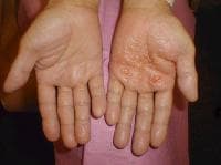
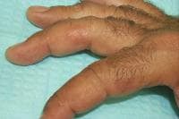
Symmetric crops of clear vesicles and/or bullae on the palms and lateral aspects of fingers characterize dyshidrotic eczema (see the images below). Feet, soles, and the lateral aspects of toes also may be affected.
Tense vesicles and bullae on the palm. Courtesy of Norman Minars, MD, University of Miami, Department of Dermatology & Cutaneous Surgery.
Small tense vesicles on the fingers.
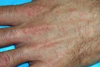
Small, discrete, coalesced vesicles on the dorsal hand.
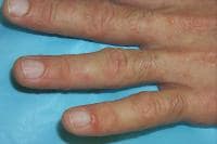
Small, discrete, coalesced vesicles on the fingers.
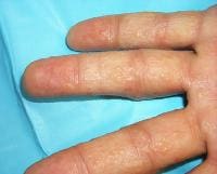
In mildly affected patients, vesicles are present only on the lateral aspects of the fingers and, occasionally, involve feet and toes (see image below).
Small discrete vesicles of the lateral fingers.
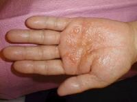
Vesicles are deep seated with a tapiocalike appearance, without surrounding erythema. They may become large, form bullae, and become confluent. Vesicles typically resolve without rupturing, followed by desquamation. See image below.
Close-up view of tense vesicles and bullae of the palm. Courtesy of Norman Minars, MD, University of Miami, Department of Dermatology & Cutaneous Surgery.
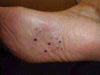
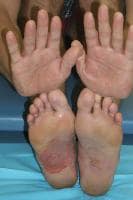
Hands are involved solely in 80% of patients, feet solely in 10%), and both hands and feet are involved in 10% of patients. See the images below.
Discrete yellow pustules on the sole of the foot. Courtesy of Norman Minars, MD, University of Miami, Department of Dermatology & Cutaneous Surgery.
Palms and soles of a patient with a dyshidrosis flare. The patient unroofed a large bulla on the right sole.
With long-standing disease, patients' fingernails may reveal dystrophic changes (eg, irregular transverse ridging, pitting, thickening, discoloration). Interdigital maceration and desquamation of the interdigital spaces often are present, despite the possible absence of a dermatophyte infection. Vesicles and/or bullae may become infected secondarily, and pustular lesions may be present. Cellulitis and lymphangitis may develop.
The Dyshidrotic Eczema Area and Severity Index was developed based on severity grades for the number of vesicles per square centimeter, erythema, desquamation, itch, and the extent of affected areas.12 The index was found to be a simple standardized method for assessing the condition and was used to assess disease severity and treatment effectiveness in 2 clinical studies. Further evaluation with larger patient groups is needed.
Causes
The cause of dyshidrotic eczema is unknown. The condition often appears related to other skin diseases (eg, atopic dermatitis, contact dermatitis, allergy to ingested metals, dermatophyte infection, bacterial infection, environmental or emotional stress). Several factors may participate in causing dyshidrotic eczema, as follows:
- Genetic factors: Monozygotic twins have been affected simultaneously by dyshidrotic eczema. The pompholyx gene has been mapped to band 18q22.1-18q22.3, in the autosomal dominant form of familial pompholyx.13
- Atopy: As many as 50% of patients with dyshidrotic eczema have reportedly had personal or familial atopic diathesis (eczema, asthma, hayfever, allergic sinusitis). The serum immunoglobulin E (IgE) level frequently is increased, even in patients who do not report a personal or familial history of atopy. Occasionally, dyshidrotic eczema is the first manifestation of an atopic diathesis.
- Nickel sensitivity: This may be a significant factor in dyshidrotic eczema. Nickel sensitivity was reportedly low in some studies of dyshidrosis patients but significantly elevated in other studies. Increased nickel excretion in the urine has been reported during exacerbations of pompholyx. Ingested metals have been found to provoke exacerbations of pompholyx in some patients.
- Low-nickel diets: These have reportedly decreased the frequency and severity of pompholyx flares. A high palmoplantar perspiration rate has been suggested to result in a local concentration of metal salts that may provoke the vesicular reaction. Contact allergy has been documented in 30% of patients with dyshidrotic eczema.
- Cobalt sensitivity: The oral ingestion of cobalt manifests systemic allergic dermatitis as dyshidrotic eczema less frequently than the oral ingestion of nickel. Much more common is the simultaneous occurrence nickel and cobalt allergy seen in 25% of nickel-sensitive patients developing pompholyx. In these cases, the eczema is usually more severe. When suspected as the cause of the dyshidrotic eczema, high oral ingestion of cobalt should be taken in consideration, regardless of the patch test results.3
- Low-cobalt diet: A point-based diet has been proposed to help patients limit cobalt ingestion and to keep the serum level below the threshold for developing flares, which is approximately less than 12 mcg/d. This diet has demonstrated higher compliance than an avoidance diet list. In addition, this diet also reduces the amount of nickel consumed.3
- Exposure to sensitizing chemicals or metals: Dyshidrotic eczema outbreaks are sometimes associated with exposure to sensitizing chemicals or metals (eg, chromium, cobalt, carba mix, fragrance mix, diaminodiphenylmethane, dichromates, benzoisothiazolones, paraphenylenediamine, perfumes, fragrances, balsam of Peru, Primula plant).
- Id reaction: Controversy surrounds the possible existence of an id reaction, which is considered to be a distant dermatophyte infection (tinea pedis, kerion of scalp) triggering a palmar pompholyx reaction (also termed pompholyx dermatophytid).
- Fungal infection: Pompholyx occasionally resolves when a tinea pedis infection is treated, then relapses when the fungal infection recurs, supporting the existence of this reaction pattern. Of patients who have a vesicular reaction to intradermal trichophytin testing, less than one third have experienced a resolution of pompholyx after treatment with antifungal agents.
- Emotional stress: This is a possible factor in dyshidrotic eczema. Many patients report recurrences of pompholyx during stressful periods. Improvement of dyshidrotic eczema using biofeedback techniques for stress reduction supports this hypothesis.
- UV-A light: A group of 5 patients with pompholyx developed typical lesions morphologically and histologically consistent with a vesicular dermatitis after a provocation with long-wavelength UV-A light. Of interest, UV-B phototherapy and photochemotherapy are well-known efficient therapies for pompholyx. Further studies ruled out contact dermatitis, polymorphic light eruption, and heat as the culprit of the lesions in these patients, confirming that the reaction was a true photosensitivity rather than a photoaggravation.14 Pompholyx caused by exposure to UV-A may be considered a variation of seasonal (summer) pompholyx.
- Other factors: Isolated reports describe other possible causative factors, such as aspirin ingestion, oral contraceptives, cigarette smoking, and implanted metals, among others. A 3-year prospective study of the causes of dyshidrotic eczema (pompholyx) in 120 patients found causes of pompholyx related to contact exposure (67.5%), including cosmetic products (31.7%) and metals (16.7%); interdigital-plantar intertrigo (10%); and internal causes (6.7%), with an additional 15% with undiagnosed (idiopathic) causes, probably related to atopic factors.15 Specifically, note the following:
- Contact allergy was found in 89 (74.2%) of 120 of the patients. The most frequent allergens were nickel, shower gel, chromium, fragrance, shampoo, and balsam of Peru. Less frequent allergens were lanolin, cobalt, thiuram, lauryl sulfate, fresh tobacco, p -phenylenediamine (PPD), formaldehyde, parabens, and octyl gallate. In 97 of 193 positive patch test results, correlation existed between the application of the agent and pompholyx recurrence. The relevance of the analysis was confirmed in 81 (67.5%) of 120 patients. In summary, the most frequent causes of pompholyx related to contact with substances were hygiene product intolerance (46.7%), metal allergy (25%), and others (28.3%).
- Intertrigo occurred in 19 (15.8%) of 120 patients. Of those, 80% presented with dermatophytosis and 20% presented with candidiasis. After 3 weeks of antifungal therapy, 6 of 19 patients remained symptomatic for pompholyx.
- With regard to internal causes, 30 patients presented with a positive patch test result for metals, but only 2 presented with exacerbations of the lesions after a challenge test.
- Of 58 patients with a history of smoking tobacco, 5 presented with a positive reaction, and 2 of those reactions were considered relevant.
- Drug allergy was determined to be the causative agent in 3 patients (amoxicillin in 2 and intravenous immunoglobulin in 1).
- Food-related pompholyx was detected in 4 patients, and after a challenge test, reactivation occurred in 3 patients (2 for paprika and 1 for orange juice).
It's a nice article. Everyone should read. Thanks for sharing. For more amazing information you may get from this link Eczema
जवाब देंहटाएं